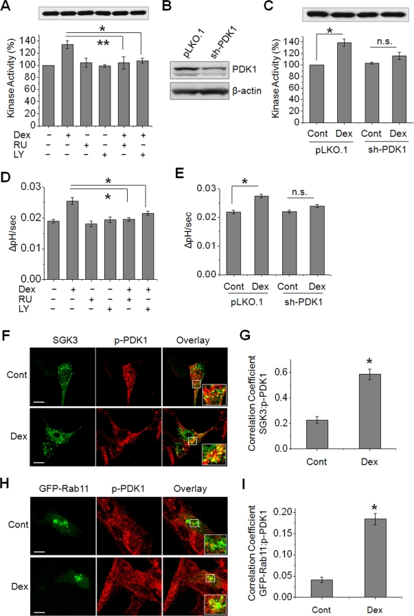
FIGURE 8:
SGK3-mediated activation of NHE3 is dependent on PI3K and PDK1. (A) PS120/NHE3V/HA-SGK3 cells were pretreated with 1 μM RU or 20 μM LY prior to treatment with 1 μM Dex or carrier for 15 min. SGK3 protein was purified by immunoprecipitation, and the kinase activity was determined by TR-FRET. n = 3. (B) PS120/NHE3V/HA-SGK3 cells were stably transfected with sh-PDK1 or pLKO.1, and the expression level of PDK1 was determined by Western blotting using an anti-PDK1 antibody. (C) PS120/NHE3V/HA-SGK3 cells transfected with sh-PDK1 or pLKO.1 were treated with 1 μM Dex or carrier for 15 min. SGK3 protein was purified, and kinase activity was determined. n = 3. (D) NHE3 activities were determined in PS120/NHE3V/HA-SGK3 cells pretreated with RU or LY prior to treatment with Dex. (E) NHE3 activities in PS120/NHE3V/HA-SGK3 cells transfected with sh-PDK1 or pLKO.1 are shown. (A–E) n.s., not significant. *, p < 0.01 and **, p < 0.05, compared with respective controls. n = 6. (F) PS120/NHE3V cells expressing HA-SGK3 were serum-starved and treated with Dex or carrier for 15 min. Colocalization of SGK3 (green) and phospho-PDK1(p-PDK1, red) was determined using anti-HA and anti-p-PDK1 antibodies, respectively. (G) The graph represents Pearson's coefficient of SGK3 and p-PDK1 colocalization from 10 independent fields of cells. *, p < 0.01 compared with the control. (H) Colocalization of GFP-Rab11 (green) and p-PDK1 (red) in PS120/NHE3V cells treated with or without Dex for 15 min is shown. Scale bar: 10 μm. Insets show high magnification of merged signals. (I) Pearson's coefficient of GFP-Rab11 and p-PDK1 colocalization from 10 independent fields of cells. *, p < 0.01 compared with the control.

