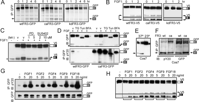
FIGURE 2:
FGF1-induced cleavage requires FGFR3 activation. (A–H) Arrow, cleaved FR3; asterisk, nonspecific band. (A) Pulse-chase analysis of T-Rex 293 cell lines stably expressing wt, ca, or kdFR3-GFP in growth media, immunoprecipitated for GFP epitope. (B–D) Western blots of wtFR3-expressing T-Rex 293 cells, cultured in DMEM/FBS(–) media in the presence or absence of FGF1 and probed for C-terminal V5 epitope, unless stated otherwise. (B) Serum-starved cells activated with FGF1 for the times indicated. Bottom, longer exposure to show that kd receptor is not cleaved. (C) Cells treated 8 h with or without tet/FGF1, ± PD173074 (PD), or SU5402 as indicated. v, vehicle (dimethyl sulfoxide [DMSO]). (D) Cells cultured 8 h in the presence or absence of inhibitor as indicated. BFA, 6 μg/ml brefeldin A; TG, 1 mg/ml thapsigarin; Tun, 1 μg/ml tunicamycin; v, DMSO. Top, 1/10 WCL, probed for GFP. Bottom, IP GFP, IB phosphotyrosine (pY20). wtFR3, left; caFR3, right. (E) Western blot of Cos7 cells stably expressing wtFR3-GFP cultured 5 h at 37°C (left) or 23°C (right), probed for GFP. (F) Western blot of Cos7 cells stably expressing wt or caFR3-GFP cultured under growth conditions, immunoprecipitated for GFP and probed for phosphotyrosine (pY20, left). WCL 1/10 probed for GFP (right). (G) Serum-starved wtFR3-GFP expressing T-Rex 293 cells, activated 5 min with the growth factors indicated. Equal micrograms of lysate were immunoprecipitated for GFP and probed for phosphotyrosine (pY20). WCL 1/10 was probed for GFP (bottom, IB GFP). (H) T-Rex 293 cells expressing wtFR3 were tet induced for 8 h in DMEM/FBS(−) media containing the FGF species indicated. Western blot of lysates probed with V5 antibodies. Bottom, longer exposure to show relative amounts of cleaved FR3.

