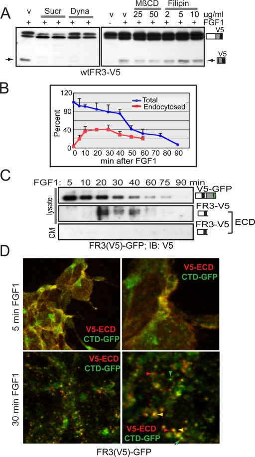
FIGURE 3:
Endocytosis is required for FGF1-induced cleavage of FGFR3. (A) Western blot of lysates from wtFR3-V5–expressing T-Rex 293 cells induced 3 h ± FGF1 before treating an additional 5 h with drug or DMSO (v). Dyna, 67, 80 μM dynasore; MβCD, methyl-β-cyclodextrin; Sucr, 0.4 M sucrose. (B) Graphical analysis of three independent experiments plotting loss of intact cell surface biotinylated receptor (blue line) and endocytosis of intact, cell surface biotinylated, glutathione-resistant receptor (red line). SDs are given as error bars. (C) FR3(V5)-GFP expressing cells were serum starved, cell surface biotinylated, and then treated with FGF1 for the times indicated. Equal micrograms of lysate or all the culture media (CM) was affinity purified using NeutrAvidin Gel. Western blot using equal volumes of purified lysate or one half of purified CM was probed for N-terminal V5 epitope (ECD). ECD was detected by overexposing blots. (D) Serum-starved FR3(V5)-GFP–expressing cells were surface labeled with V5 antibodies, then stimulated with FGF1 to promote receptor internalization. Cells were fixed, permeabilized, probed with secondary antibodies (Alexa 543) to the endocytosed V5 epitope, and imaged by confocal microscopy. Right, higher-magnification image of left. Green, C-terminal GFP; red, endocytosed ECD-V5. Green arrowheads, GFP-containing vesicles; red arrowheads, ECD-containing vesicles; yellow arrowhead, colocalized ECD/CTD.

