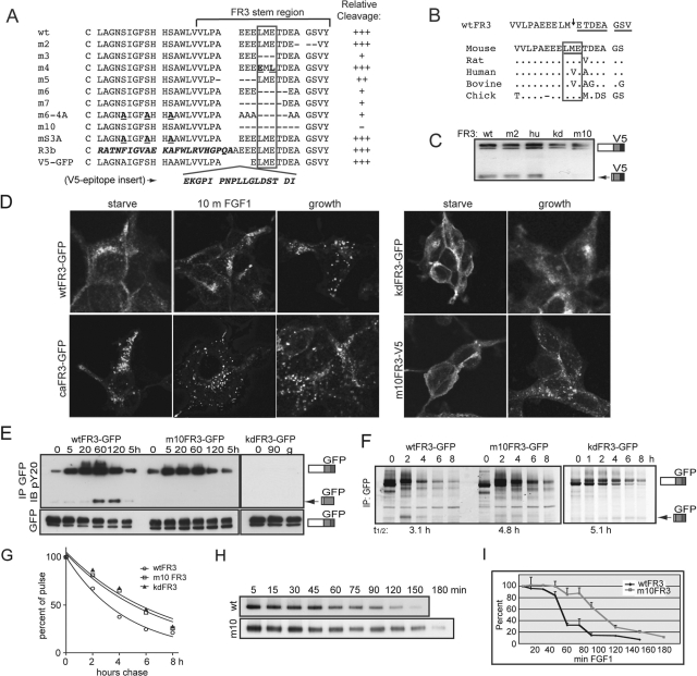
FIGURE 5:
Disrupting cleavage alters the half-life, but not the trafficking, of FGFR3. (A–I) T-Rex 293 stable cell lines. Arrow, cleaved FR3. (A) Stem region sequence of mutants (m) generated and relative degree of cleavage, averaged from at least three independent experiments. Cell lines were tet-induced 8 h in the presence of FGF1; cleavage was assessed by Western blot using V5 or GFP epitope. +++, wt cleavage; ++, reduced cleavage; +, minimal cleavage; −, no cleavage detected. Putative cleavage region is boxed. Mutated residues are shown in bold; FR3b-specific sequence and V5 epitope insertion are shown in bold and italicized. (B) Top, cleavage site determined by N-terminal sequencing of gel-purified 72-kDa fragment. Arrow, cleavage site. Sequenced residues are underlined. Bottom, homology of cleavage region between species. Residues surrounding cleavage site are boxed; distinct amino acids are listed. (C) Representative Western blot of various stable FR3-V5 cell lines probed for V5 epitope. hu, human stem region. (D) Confocal images of wt and mutant FR3 following serum starvation, 10 min FGF1 addition, or cultured in growth media. GFP imaged directly; V5 probed with V5 antibody and Alexa 488 secondary antibody. (E) Cells expressing wt and mutant FR3-GFP were serum starved and then treated with FGF1 for the times indicated. Equal micrograms of lysates were immunoprecipitated for GFP and probed for phosphotyrosine (pY20). (F) Cells expressing wt, m10, and kdFR3-GFP were subject to pulse-chase analysis in the presence of FGF1 and immunoprecipitated for GFP. Arrow, cleaved FR3. T1/2, half-life of intact receptor calculated using GraphPad (La Jolla, CA). (G) Graphical depiction of the half-life for intact wt, m10, and kdFR3. (H) Cells expressing wt or m10FR3-V5 were serum starved, cell surface biotinylated, and stimulated with FGF1 for the times indicated. Equal micrograms of lysate were immunoprecipitated using NeutrAvidin Gel; the stability of the intact receptor was assessed by Western blot using the V5 epitope. (I) Graphical analysis of three independent experiments assaying loss of intact cell surface biotinylated receptor. SDs are shown as error bars.

