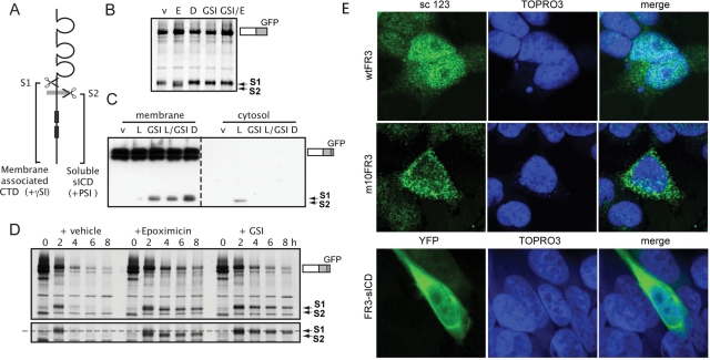
FIGURE 6:
FGFR3 is cleaved by γ-secretase. (A) Cartoon of cleavage sites (S1, putative cathepsin; S2, γ-secretase) and their subcellular localization. γSI, γ-secretase inhibitor; PSI, proteasome inhibitor. (A) Cells expressing wtFR3-GFP were pulse labeled in the presence of FGF1 and then chased 2.5 h in the presence of inhibitors. D, 25 μM DAPT; E, 1 μM epoxomicin; GSI, 5 μM compound E; v, DMSO. (C) Western blot of cells expressing wtFR3-GFP, cultured 5 h in the presence of inhibitors, subject to subcellular fractionation, and probed for C-terminal GFP tag. Left, membrane-associated proteins; right, cytosolic proteins. L, 10 μM lactacystin. (D) Cells expressing wtFR3-GFP were pulse labeled and then chased in the presence of FGF1 plus inhibitors. Bottom, longer exposure to show the size shift of the cleaved fragment. Dashed line, S1-cleaved FR3. (E) Confocal images of cells transiently overexpressing untagged wtFR3 (top), m10FR3 (middle), or YFP-tagged FR3-sICD (bottom, YFP-sICD). sc 123, C-terminal anti-FGFR3 antibody (green); YFP pseudo-colored green; TOPRO3, nuclear stain (blue).

