Abstract
Purpose
To use computer navigation software to investigate the specific origins of the hip capsuloligamentous complex.
Methods
Six fresh frozen cadaver hips were anatomically landmarked utilizing a three-dimensional computer navigation system. The acetabular origins of the iliofemoral, pubofemoral, and ischiofemoral ligaments were statically digitized. Computer software was used to create a 180° (6:00) meridian line positioned over the midpoint of the acetabular notch, and to present the results in a clocklike manner in hours and minutes (00:00) and also degrees relative to the 12 o’clock position.
Results
The iliofemoral ligament origin starts at 17° (±31°) from the 12 o’clock position, or 12:35 (±1:02) in hours and minutes, and ends at 69° (±13°) or 2:18 (±0:25), spanning a mean distance of 52° (±19°). The ischiofemoral ligament has the broadest origin, starting at 262° (±12°) or 8:44 (±0:24), and ending at 353° (±17°) or 11:45 (±0:14), spanning a mean distance of 90° (±6°). The pubofemoral ligament origin is the smallest, starting at 121° (±5°) or 4:02 (±0:11), and ending at 163° (±9°) or 5:27 (±0:18), spanning a mean distance of 42° (±5°). The iliofemoral ligament origin demonstrates the greatest anatomic variability with regards to its location and its size (p = 0.002).
Conclusion
This study demonstrates that there is significant variability in the size and location of the iliofemoral ligament origin versus the pubofemoral and ischiofemoral ligaments.
Level of Evidence
Level IV anatomic cadaveric study. See the guidelines online for a complete description of level of evidence.
Keywords: hip ligaments, computer navigation, hip capsule, hip arthroscopy
Introduction
The unique anatomy of the human hip allows for functional mobility, yet provides the necessary constraints to maintain stability [6]. In addition to the hip’s inherent bony constraints, further stability is imparted by a strong sleeve of fibrous capsule that encloses the hip joint, extending from the bony ridge of the acetabulum to the proximal femur. This capsule is intricately reinforced with a series of three longitudinally oriented ligaments: the iliofemoral, ischiofemoral, and pubofemoral ligaments, in addition to a circularly oriented layer known as the zona orbicularis [11]. Prior research concerning these ligaments has focused primarily on their biomechanical properties and contributions to stability, with numerous studies investigating the forces at failure of each respective ligament [1, 4, 5, 9].
Although a multitude of research has been performed analyzing the interactions that maintain biomechanical mobility and stability of the hip, surprisingly little information exists detailing the anatomic characteristics of these ligaments [3]. In addition, with the advent of arthroscopic management of intra-articular hip pathology, and new techniques for hip preservation, there has been an increase in the use of capsulotomies to improve exposure during arthroscopy, but little data exists regarding the exact structures cut (capsule, ligaments, or both), and the resulting effect on postoperative stability. Understanding the exact origins of the hip ligaments could further elucidate their specific characteristics, and possibly lead to less traumatic arthroscopic approaches and techniques.
Therefore, the aims of this study were to describe the specific acetabular origins of the iliofemoral, ischiofemoral, and pubofemoral ligaments utilizing computer navigation software. This information is presented in a clocklike manner to increase the ease of conceptualization during arthroscopic hip surgery. In addition, we wished to describe any variability in the size and location of the acetabular origins of each ligament detected in our specimens.
Materials and Methods
Three fresh frozen cadaveric pelvises providing a total of six hips (three right, three left) were used in this IRB-approved study. Prior to formal testing of the specimens, a pilot study utilizing a single, fresh-frozen human cadaver hip was performed to test the experimental protocol.
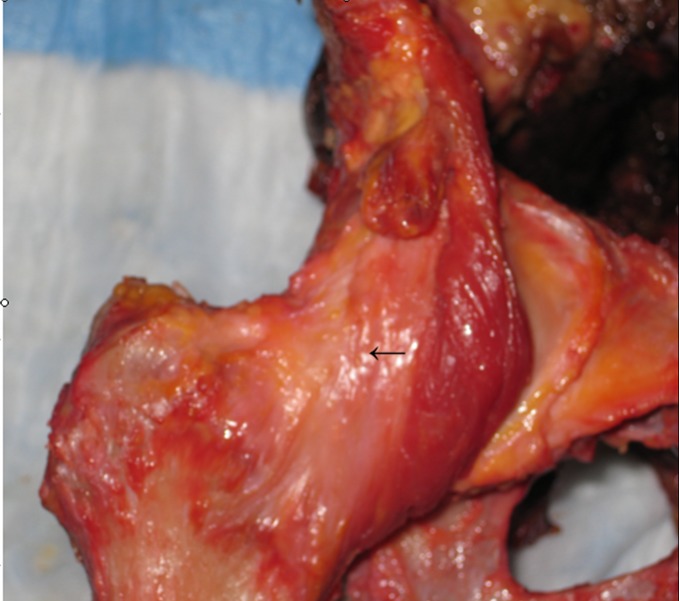
All specimens were thawed at room temperature for at least 24 h prior to testing. A radiographic and manual examination of each hip and pelvis was performed to ensure that no occult pathologies were present. If any signs of arthrosis, abnormal femoral head–neck offset, acetabular retroversion, acetabular overcoverage, decreased mobility, surgical interventions, or crepitation was found, the specimen was excluded from this project. All of the tissue surrounding the hip was carefully dissected for full identification of the capsuloligamentous structures, including the iliofemoral, ischiofemoral, and pubofemoral ligaments (Fig. 1).
Fig. 1.
Photograph of the right hip of a cadaveric specimen demonstrating the anterior structures of the hip capsuloligamentous complex after surgical dissection. The arrow is pointing to the fibers of the iliofemoral ligament
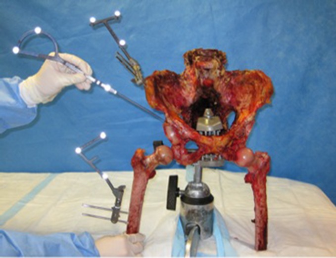
Each intact pelvis was mounted to a table stand by rigidly clamping the pelvis. Two Steinmann pins were drilled into the midshaft of the femoral diaphysis, and two in the iliac crest, approximately 2 cm behind the anterior superior iliac spine. Infrared reflector pads were attached to each metal rod complex via a metal clamp, in order to establish reference points on the femur and pelvis for use with a three-dimensional computer navigation software system (Praxim, La Tronche, France) (Fig. 2). The computer navigation system used was an imageless system in which anatomical landmarking and bone morphing is performed using a three-space digitizer to create a three-dimensional computer replica of the specimen’s anatomy. The leg was then manually cycled through a full range of passive motion to ascertain the center of the femoral head and the center of rotation of the hip joint. With the sensory markers left in place, a three-space digitizer was then calibrated. Bony landmarks of the hip joint, including the most anterior portion of the anterior inferior iliac spine (AIIS), were then recorded by the navigation software.
Fig. 2.

Demonstration of the experimental set-up with the cadaveric pelvis rigidly mounted to a table stand, and infrared reflector pads attached to both the ilium and the femur to establish reference points for use with the computer navigation system. The capsuloligamentous structures have already been removed and the hip has been disarticulated in this specimen
The acetabular origins of the ischiofemoral, iliofemoral, and pubofemoral ligaments were then statically digitized, utilizing 11 points of reference for the origin of the iliofemoral ligament, 7 points of reference for the pubofemoral ligament, and 12 points of reference for the ischiofemoral ligament. The femur was then disarticulated from the pelvis, and the intra-articular surface geometry was mapped. Morphing the proximal acetabulum and capsuloligamentous origins with a three-space digitizer reproduces a three-dimensional computer replica of these structures that is accurate to within 1 mm and 1° [10]. The accuracy of the acquisition was verified by correlating the location of the digitizer, depicted on the video display, with the location on the physical specimen.
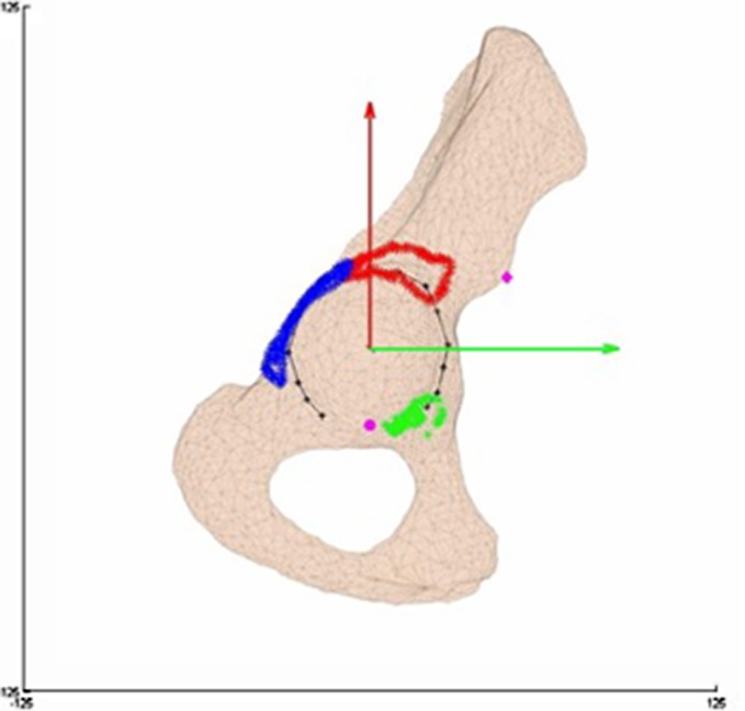
After the anatomic acquisition was complete, reference points and the center of the acetabulum were utilized to determine the origins of the iliofemoral, ischiofemoral, and pubofemoral ligaments relative to a 180° (6:00) meridian line positioned over the midpoint of the acetabular notch, as described by Köhnlein et al. [7]. The 90° latitude line defined the equatorial line of a hemisphere. This was performed utilizing Matlab software (The Mathworks™ Inc., Novi, MI). The data sets obtained from the computer navigation system for each specimen were incorporated into the Matlab generated coordinate system in the same orientation, to maintain consistency among the specimens during data analysis. The origins of the iliofemoral, ischiofemoral, and pubofemoral ligaments, in addition to the location of the AIIS, were recorded in a clocklike manner in hours and minutes (00:00), and also degrees relative to the 12 o’clock position (°) (Fig. 3). For standardization, all data on left-sided specimens were mirrored along the 180° meridian line and recorded as right-sided acetabula, as described by Köhnlein et al. [7].
Fig. 3.
Example of the image created after digitization of the iliofemoral, ischiofemoral, and pubofemoral ligament origins and the anterior inferior iliac spine (AIIS), with the axes of the clockface superimposed. Red arrow, 12 o’clock; green arrow, 3 o’clock; pink diamond, AIIS; pink circle, middle of the acetabular notch; red outline, iliofemoral origin; blue outline, ischiofemoral origin; green arrow, pubofemoral origin
All data analyses were performed using SPSS software (SPSS Inc., Chicago, IL). Calculations were performed to determine the average deviation from the mean, with respect to the start and end points of each ligament’s origin, and the actual length of each ligamentous origin. Two-tailed Student’s t-tests were used to determine if there was a statistically significant difference in the deviation from the mean with regards to the length of each respective ligamentous origin. P values <0.05 were considered statistically significant. The standard deviations of all numerical data are presented in parentheses (±SD).
Results
All three ligaments have well-defined acetabular origins that can be described using the orientation of a clock face. The position of the AIIS relative to the acetabular clockface occurs at the 2:08 (±0:04) position, or 64° (±2°) from the 12 o’clock position. As landmarking of the AIIS was performed with the acquisition of one data point in our protocol (the most anterior aspect of the AIIS), no data is present with regards to the start and end point of the AIIS, and thus its variability. The mean midpoint of the origin of the iliofemoral ligament is located at the 1:26 (±0:43) position, as on average, the ligament’s origin begins at 12:35 (±1:02) and extends to 2:18 (±0:25). In degrees, the ligament’s origin starts at 17° (±31°) from the 12 o’clock position, and ends at 69° (±13°), spanning a mean distance of 52° (±19°) around the acetabular clockface. The ischiofemoral ligament was found to have the largest origin, with a mean midpoint located at the 10:15 (±0:19) position, starting at 8:44 (±0:24) and terminating at 11:45 (±0:14). In degrees, the ligament’s origin starts at 262° (±12°) and ends at 353° (±17°) from the 12 o’clock position, spanning a mean distance of 90° (±6°). The pubofemoral ligament origin was the smallest measured, with a mean midpoint located at the 4:44 (±0:14) position. On average, the ligament’s origin begins at 4:02 (±0:11) and extends to the 5:27 (±0:18) position. In degrees, the ligament’s origin starts at 121° (±5°) and ends at 163° (±9°), spanning a mean distance of 42° (±5°).
The iliofemoral ligament had the greatest variability with regards to the starting point and end point of its origin, and overall size, when compared to the ischiofemoral and pubofemoral ligaments. The starting point of the iliofemoral ligament demonstrated an average deviation from the mean starting point of 27° (versus 10° and 5°, respectively). Therefore, on average, the starting point of the iliofemoral ligament in each specimen was 27° different than the mean starting point for all iliofemoral ligament specimens measured. In addition, the end point of the iliofemoral ligament origin also demonstrated the greatest variability when compared to the ischiofemoral and pubofemoral ligaments, demonstrating an average deviation from the mean end point of 10° (versus 6° and 7°, respectively). Subsequently, the iliofemoral ligament possessed the greatest average deviation from the mean in the overall size of the origin of the ligament versus the ischiofemoral and pubofemoral ligaments, with a value of 16° versus 5° and 3°, respectively. Therefore, the iliofemoral ligament origin demonstrated the greatest anatomic variability (p = 0.002) (Table 1).
Table 1.
Values obtained for the origins of iliofemoral, ischiofemoral, and pubofemoral ligaments
| Average starting point (°) | Average end point (°) | Average midpoint (°) | Average length of origin (°) | Average deviation of the length of the origin (°) | |
|---|---|---|---|---|---|
| Iliofemoral ligament | 17 (±31) | 69 (±13) | 43 (±22) | 52 (±19) | 16 |
| Ischiofemoral ligament | 262 (±12) | 353 (±17) | 307 (±10) | 90 (±6) | 3 |
| Pubofemoral ligament | 121 (±5) | 163 (±9) | 142 (±7) | 42 (±5) | 5 |
Data are presented as degrees ± standard deviation relative to the 12 o’clock position on the acetabular clockface. The iliofemoral ligament demonstrated the greatest average deviation of the length of its origin, as on average, the length for each specimen varied 16° from the mean value for the length of all iliofemoral ligaments measured. This difference was statistically significant when compared to the ischiofemoral and pubofemoral ligaments (p = 0.002)
Discussion
The aims of this study were to describe the distinct acetabular origins of the iliofemoral, ischiofemoral, and pubofemoral ligaments, and present this information in terms of a clockface for ease of intraoperative conceptualization. In addition, any variability seen among our specimens in the size and location of the acetabular origins of each ligament would be presented. As noted above, the iliofemoral, ischiofemoral, and pubofemoral ligaments were found to have well defined acetabular origins amenable to being described using the orientation of a clockface. With regards to anatomic variability among the specimens tested, the iliofemoral ligament demonstrated significant variability in both its location and size, when compared to the ischiofemoral and pubofemoral ligaments.
There were several limitations to this study, including the small sample size presented. However, despite the small sample size, a statistically significant difference was detected with regards to the anatomic variability of the iliofemoral ligament origin when compared to the ischiofemoral and pubofemoral ligaments. A second limitation is that the accuracy of anatomic landmarking when using the imageless computer navigation system may have varied among the specimens tested. However, during anatomic landmarking and data acquisition, the accuracy of each point was verified by correlating the location of the digitizer, depicted on the video display, with the location on the physical specimen. In addition, data regarding the ligament insertions into the femur would have provided useful information regarding the orientation of the ligaments, and the possibility of setting guidelines for a less traumatic capsulotomy.
With regards to origin size, the ischiofemoral ligament was found to have the largest origin, spanning a mean distance of 90° (±6°) around the acetabular clockface, while the pubofemoral ligament origin was the smallest, spanning a mean distance of 42° (±5°). The position of the AIIS was consistently found to be at the 2:08 position of the acetabular clockface, with the acetabular notch being at a standardized location of 6:00 based on our experimental protocol. To our knowledge, no prior study has used computer navigation to either determine the specific acetabular origins of these ligaments, or present this information relative to a clockface. Köhnlein et al. [7] used an acetabular clockface to define the acetabular rim profile and acetabular morphology in cadaveric specimens, however the soft tissue anatomy was not assessed.
In our study, the acetabular origin of the iliofemoral ligament was found to be highly variable both in its location and overall size, when compared to the ischiofemoral and pubofemoral ligaments. The significance of this variation is unknown, and has not been previously described. Similar variations in the origin of the anterior and posterior bands of the inferior glenohumeral ligament complex of the shoulder have previously been demonstrated by O’Brien et al. [8]. DePalma et al. [2] postulated that these variations either represent an inherent anatomical variability, or possibly a functional adaptation to individual joint geometry, which may also be applicable to the variability seen in the iliofemoral ligament origin. Therefore, further investigations are required to determine the clinical significance of this finding in the hip, and perhaps its effect on mechanical stability.
In conclusion, this study describes the distinct acetabular origins of the iliofemoral, ischiofemoral, and pubofemoral ligaments relative to an acetabular clockface, and demonstrates that the iliofemoral ligament origin has significant anatomic variability relative to the ischiofemoral and pubofemoral ligaments. Further studies are currently underway utilizing microcomputed tomography to assess if there are portions of the ligament that are consistently less robust, and thus less crucial to overall ligament stability, which may represent a more ideal location for a less traumatic capsulotomy.
Footnotes
Each author certifies that he or she has no commercial associations (e.g., consultancies, stock ownership, equity interest, patent/licensing arrangements, etc.) that might pose a conflict of interest in connection with the submitted article.
Each author certifies that his or her institution has approved the reporting of this study, that all investigations were conducted in conformity with ethical principles of research.
References
- 1.Crowninshield RD, Johnston RC, Andrews JG, Brand RA. A biomechanical investigation of the human hip. J Biomech. 1978;11:75–85. doi: 10.1016/0021-9290(78)90045-3. [DOI] [PubMed] [Google Scholar]
- 2.DePalma AF, Callery G, Bennett GA. Variational anatomy and degenerative lesions of the shoulder joint. Instr Course Lect. 6. 1949:255–281.
- 3.Fuss FK, Bacher A. New aspects of the morphology and function of the human hip joint ligaments. Am J Anat. 1991;192:1–13. doi: 10.1002/aja.1001920102. [DOI] [PubMed] [Google Scholar]
- 4.Hewitt J, Guilak F, Glisson R, Vail TP. Regional material properties of the human hip joint capsule ligaments. J Orthop Res. 2001;19:359–364. doi: 10.1016/S0736-0266(00)00035-8. [DOI] [PubMed] [Google Scholar]
- 5.Hewitt JD, Glisson RR, Guilak F, Vail TP. The mechanical properties of the human hip capsule ligaments. J Arthroplasty. 2002;17:82–89. doi: 10.1054/arth.2002.27674. [DOI] [PubMed] [Google Scholar]
- 6.Hughes PE, Hsu JC, Matava MJ. Hip anatomy and biomechanics in the athlete. Sports Med Arthrosc Rev. 2002;10:103–114. doi: 10.1097/00132585-200210020-00002. [DOI] [Google Scholar]
- 7.Köhnlein W, Ganz R, Impellizzeri FM, Leunig M. Acetabular morphology: implications for joint-preserving surgery. Clin Orthop Relat Res. 2009;467:682–691. doi: 10.1007/s11999-008-0682-9. [DOI] [PMC free article] [PubMed] [Google Scholar]
- 8.O'Brien SJ, Neves MC, Arnoczky SP, Rozbruck SR, Dicarlo EF, Warren RF, Schwartz R, Wickiewicz TL. The anatomy and histology of the inferior glenohumeral ligament complex of the shoulder. Am J Sports Med. 1990;18:449–456. doi: 10.1177/036354659001800501. [DOI] [PubMed] [Google Scholar]
- 9.Stewart KJ, Edmonds-Wilson RH, Brand RA, Brown TD. Spatial distribution of hip capsule structural and material properties. J Biomech. 2002;35:1491–1498. doi: 10.1016/S0021-9290(02)00091-X. [DOI] [PubMed] [Google Scholar]
- 10.Stindel E, Gil D, Briard JL, Merloz P, Dubrana F, Lefevre C. Detection of the center of the hip joint in computer-assisted surgery: An evaluation study of the surgetics algorithm. Comput Aided Surg. 2005;10:133–139. doi: 10.3109/10929080500229975. [DOI] [PubMed] [Google Scholar]
- 11.Warwick R, Williams PL. Gray's Anatomy. 35. Philadelphia, PA: Saunders; 1973. [Google Scholar]


