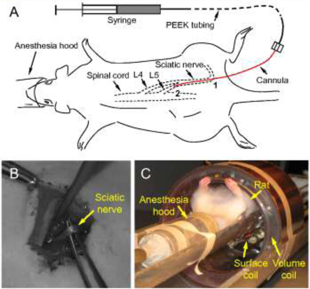
Figure 2.
(A) Schematic of sciatic nerve infusion. Point 1 is the insertion point where the infusion cannula was introduced into the sciatic nerve. Point 2 is the location of the cannula tip. Cannula length from points 1 to 2 is defined as the insertion depth. (B) The isolated rat sciatic nerve. (C) The RF dual coil system with an anesthetized rat used to collect MRI data.

