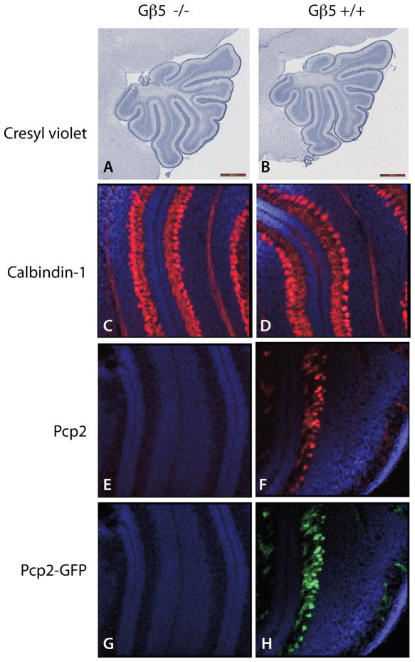
Figure 3. Delayed Purkinje cell maturation in the cerebellar cortex of seven-day old Gβ5-homozygous knockout mice.
(A, B) Cresyl violet staining of sagittal sections of cerebella of seven-day old Pcp2-GFP hemizygous Gβ5 KO and wild-type mouse littermates (scale bars = 500 μm). (C, D) Merged DAPI (blue) and calbindin-1-immunostaining (red) images of frozen sections of cerebella of Gβ5 KO and wild-type mouse littermates (50X). (E, F) Merged DAPI (blue) and Pcp2-immunostaining (red) images of frozen sections of cerebella of Gβ5 KO and wild-type mouse littermates (50X). (G, H) Merged DAPI (blue) and Pcp2-GFP reporter fluorescence (cyan) images of frozen sections of cerebella of Gβ5 KO and wild-type mouse littermates (50X). Results shown in A-H representative of three littermate pairs with similar findings.

