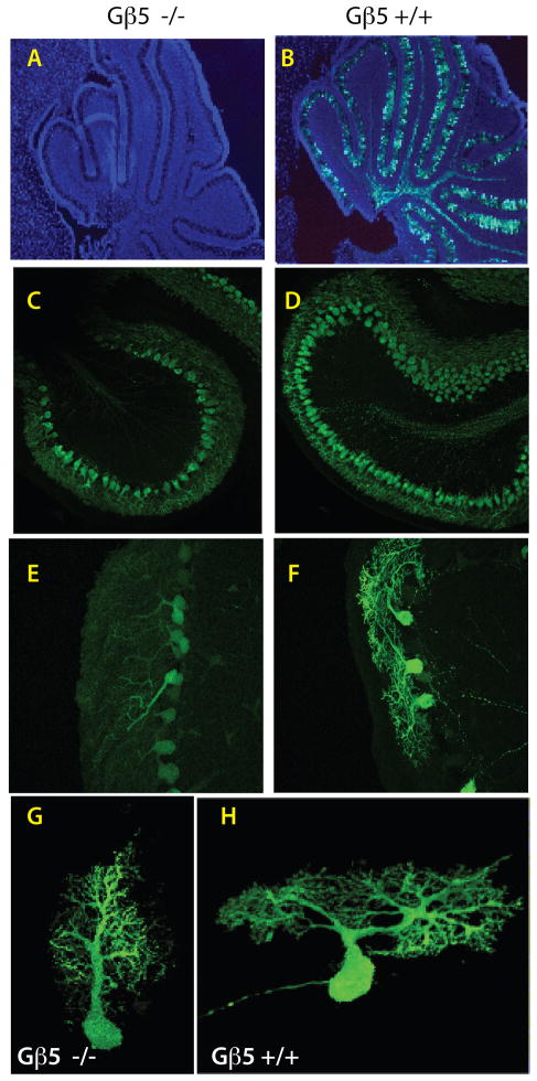
Figure 4. Delayed cerebellar development and deficient Purkinje cell maturation in Gβ5-homozygous knockout mice.
Analysis of Pcp2-GFP hemizygous Gβ5 KO (A, C, E, G) and Pcp2-GFP hemizygous Gβ5 wild-type (B, D, F, H) littermate mice. (A, B) Merged DAPI (blue) and Pcp2-GFP reporter histofluorescence (green) images from cerebellar sections harvested from seven-day old Gβ5 homozygous KO and wild-type littermates (50X). (C-H) Laser confocal analysis of Pcp2-GFP reporter histofluorescence in cerebellar sections from 10-day old mice comparing the degree of dendritic arborization in Gβ5 homozygous KO mice and their wild-type littermates. Magnification in C, D, 20 X; in E, F, 40 X. See also supplemental videos 3 and 4, corresponding to images G and H, showing 3-D images of the corresponding neurons reconstructed from a stack of confocal images acquired at 0.17 μm step size, as described in supplementary online materials and methods. Results in A-F are representative of results from three wild-type and Gnb5 KO sibling pairs.

