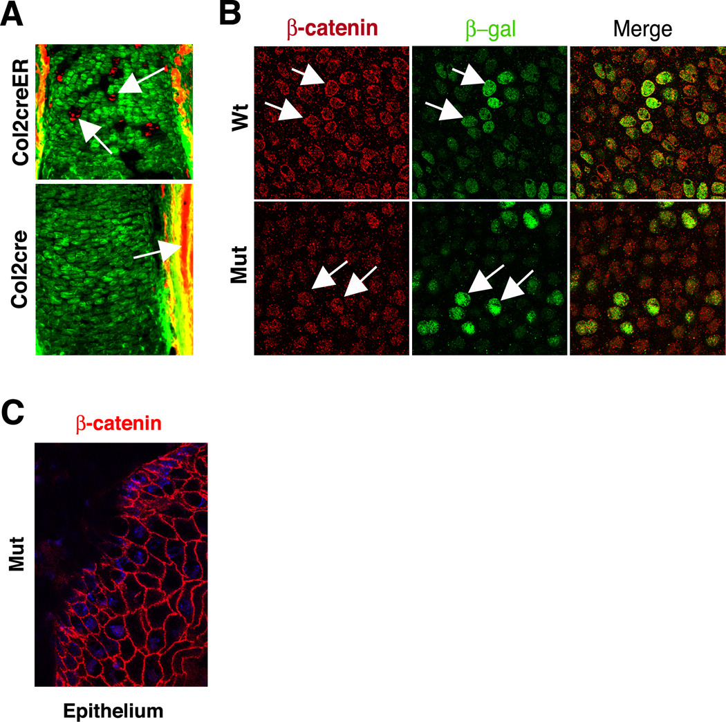
Figure 3. Efficient recombination results in the absence of β-catenin in cartilage.
(A) Cre-mediated recombination is efficient in cartilage. (Top) Epifluorescence illumination of tissue sections from embryos exposed to tamoxifen shows that the reporter locus can readily distinguish chondrocytes in which recombination occurred (green) from those in which recombination is absent (red, arrows). (Bottom) The Col2a1cre line used to generate Ctnnb1 mutants is highly efficient at promoting recombination of the reporter locus, as evidenced by the predominance of green fluorescence and the lack of red fluorescence. As expected, cells in the surrounding soft tissue lack recombination (red, arrows). (B) Immunofluorescence detection in sections of wild type cartilage demonstrates strong plasma membrane localization of β-catenin (arrows). Only some cells that express β-catenin show increased TOPGAL activity (green), as determined using an anti-β-galactosidase antibody. By contrast, Ctnnb1 mutant cartilage shows only background levels of cytoplasmic anti-β-catenin signal and not cortical localization, but retains strong TOPGAL transcription (arrows). (C) Robust expression of β-catenin (red) at the plasma membrane is found in the epithelium surrounding Ctnnb1 mutant cartilage. The nucleus is stained with DAPI (blue).

