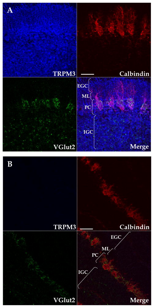
Figure 1. Pseudocolored confocal images of TRPM3 expression profile in the neonatal cerebellar cortex.
(A) TRPM3 (blue), calbindin (red) and VGlut2 (green) expression in a 2 μm-thick sagittal cerebellar confocal section at P8. Channels are shown separately and a merged image is shown in the bottom right division. Similar results were obtained in sections from 4 additional rats. (B) Competition with the immunizing peptide eliminates TRPM3 protein staining. Similar results were obtained in sections from another rat. External granule cell layer (EGC); Molecular layer (ML ), Purkinje cell layer (PC), internal granule cell layer (IGC). Scale bars: 50 μm.

