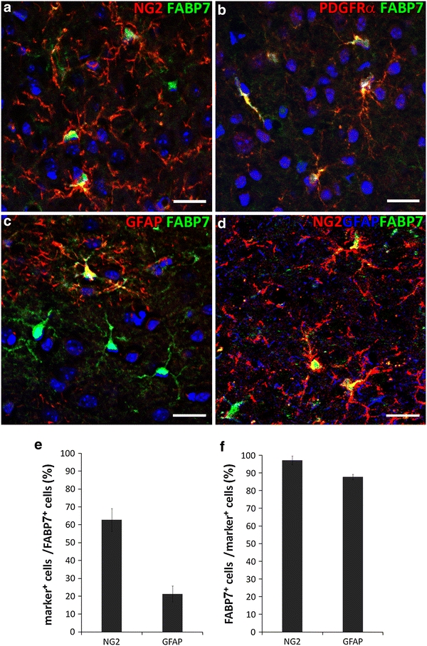
Fig. 1.

Identification of FABP7 expressing cells in the normal adult cerebral cortex. a–d Immunofluorescence micrographs showing expression of FABP7 in OPCs and astrocytes. a Expression of FABP7 (green) in NG2+ OPCs (red). b Expression of FABP7 in OPCs confirmed by colocalization of FABP7 (green) and PDGFRα (red). c Expression of FABP7 (green) in GFAP+ protoplasmic astrocyte. d Localization of FABP7 (green) in NG2+ OPCs (red) and GFAP+ astrocytes (blue). Note that the majority of FABP7+ cells are OPC. e Bar graph showing the percentage of NG2+ OPCs and GFAP+ astrocytes among total FABP7+ cells. f Bar graph showing the percentage of FABP7+ cells in NG2+ OPCs and GFAP+ astrocytes. Data in e, f are obtained from 0.1 mm2 area. Scale bars 20 μm
