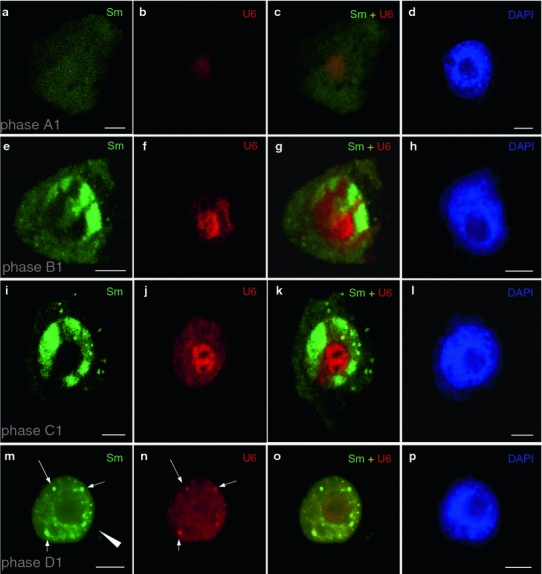
Fig. 1.

Localization of Sm proteins (mAb Y12) and U6 snRNA during premeiotic interphase. Initially, very low levels of the Sm protein were observed (a). U6 snRNA was clearly visible only in the nucleoli (b, c). In the subsequent stage, cytoplasmic bodies containing Sm proteins appeared (e). In this period, an increase in the levels of these proteins was observed in the nucleus (e, g). The amount of U6 snRNA increased considerably in the nucleoli, but a significant amount was also detected in a dispersed form in the nucleus (f). In the next stage, when cytoplasmic bodies containing Sm proteins could still be observed in the cytoplasm, high Sm protein levels were observed in the nucleoplasm and in the nuclear bodies (i, k). Colocalization of U6 in the nuclear bodies was not observed. The levels of U6 in the nucleolus were still much higher than in the nucleoplasm (j). In the last stage, only very small, single Sm bodies were visible in the cytoplasm (arrow head). In the nucleus, strong Sm protein (m) and U6 snRNA (n) signals could be observed in the nuclear bodies (arrows), whereas the signal was uniform and dispersed in the nucleoplasm (o). The corresponding DAPI images were collected using widefield fluorescence and deconvolution software (d, h, l, p). Bars 10 μm
