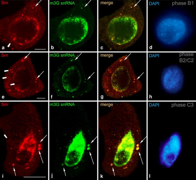
Fig. 3.

Immunolocalization of Sm (AF-ANA) proteins and m3G snRNA in the G2 (a–d), leptotene (e–h) and zygotene (i–l) phases using the double-label immunofluorescence method. Large spherical cytoplasmic bodies (arrows) containing both Sm proteins and snRNA can be observed. During this period, numerous smaller clusters of Sm proteins were also present in the cytoplasm (arrowheads), but not always together with snRNA. The corresponding DAPI images were collected using widefield fluorescence and deconvolution software (d, h, l). Bars 10 μm
