Fig. 5.

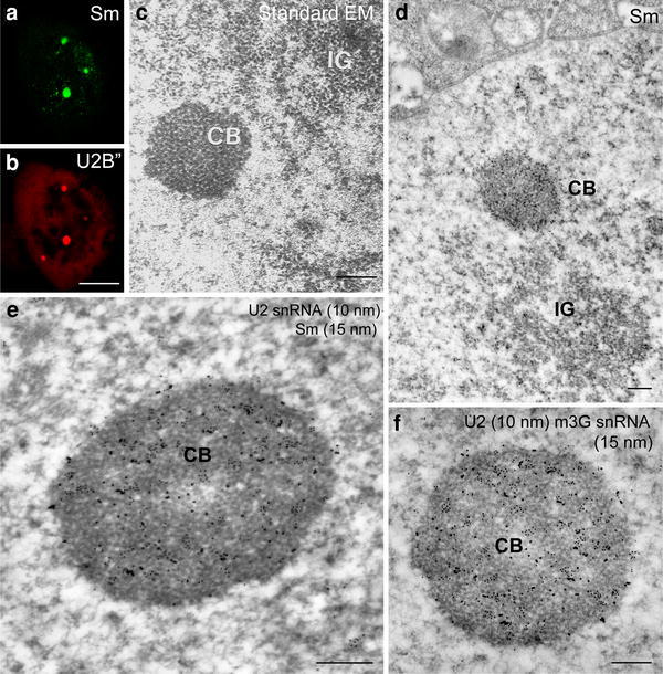
The Cajal bodies of larch microsporocytes. In the nuclei, spherical nuclear bodies were present, exhibiting a regular shape and characterized by labeling of Sm (a) and U2B proteins (b) at much higher levels than in the surrounding nucleoplasm (a, b). Bar 10 μm. c Standard electron microscopy (EM) technique. The Cajal bodies (CB) are made up of coiled fibrils of 15–25 nm in diameter, with nearby visible clusters of interchromatin granules (IG). Immunolocalization of SmD proteins (mAb Y12) (d). A cluster of intensely labeled Sm proteins was detected in the Cajal body close to the nuclear envelope and IG (d). Colocalization of U2 snRNA (10 nm gold particles) and Sm proteins (15 nm gold particles) (αSm) at the ultrastructural level (e) immunogold/high-resolution ISH method. In the Cajal bodies, labeling and colocalization of Sm proteins with U2 snRNA was very high (e). The localization of m3G snRNA (15 nm gold particles) and U2 snRNA (10 nm) labeling in the Cajal bodies is much stronger than in the surrounding nucleoplasm (f) immunogold/high-resolution ISH method. Bars 0.5 μm
