Fig. 4.

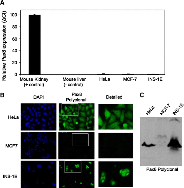
The Pax8 protein but not the transcript is detected in various cell lines. A Q-RT-PCR analysis shows nearly non-detectable expression levels of Pax8 in HeLa, MCF-7 and INS-1E cell lines. The graphic represents the relative abundance of Pax8 mRNA in the different cell lines as compared to the expression levels in kidney (arbitrarily indicated as 100). B Immunofluorescence analysis of the same cell lines shows that only MCF-7 cell line is negative for immunostaining when using the Pax8 polyclonal antibody (green) (middle panel). Counterstaining with DAPI (blue) is shown in the left panels (×400). A detailed image corresponding to an enlargement of the indicated area is showed in the right panels. C Western blot analysis of cell extracts using the polyclonal Pax8 antibody revealed the presence of an immunoreactive band of approximately 48 kD (corresponding to the molecular weight of Pax8) in all cell lines except MCF-7 cells
