Fig. 1.
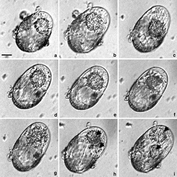
Time-lapse photographs showing the earliest stages of embryogenesis in the microspore suspension culture of Brassica napus. a–f The microspore volume enlarged and resulted in the cell emerging from the microspore parental wall at one pole. The nucleus moved from and towards the apical pole and came close to the intensively stretched sporoderm wall. g–i Chromatin condensation and slightly asymmetric mitotic division. h–i Structure with two nuclei (arrowheads). Images a–f were taken from day 7 to day 8 of culture. Cultures were continuously kept in a NLN-13 medium. Bar 20 μm

