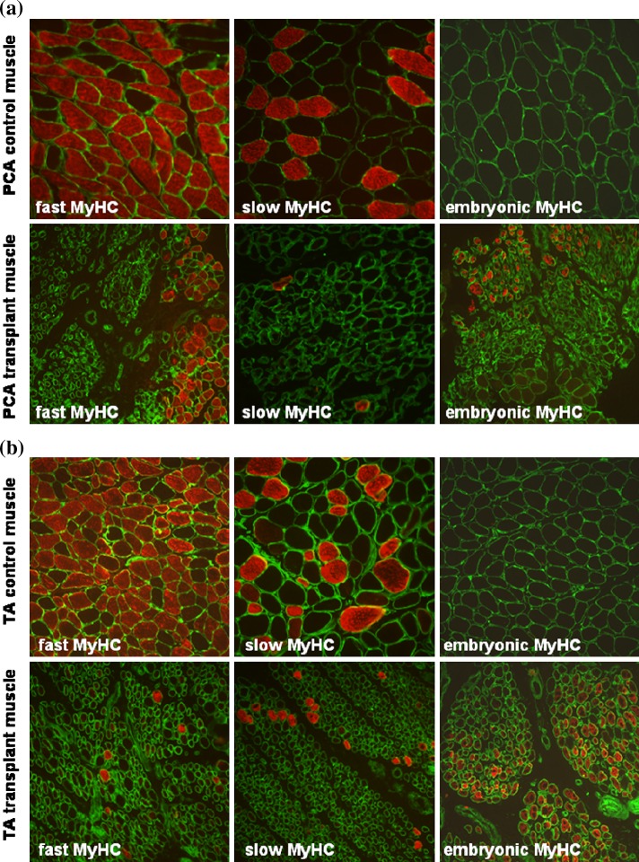
Fig. 5.
Morphology of laryngeal muscle fibres. a Posterior cricoarytenoid (PCA) muscle. Transverse sections of control PCA (top row) and transplant PCA (bottom row) muscles stained with laminin antibody (green) and either fast type, slow type or embryonic MyHC protein antibody (red). b Thyroarytenoid (TA) muscle. Transverse sections of control TA (top row) and transplant TA (bottom row) muscles stained with laminin antibody (green) and either fast type, slow type or embryonic MyHC protein antibody (red)

