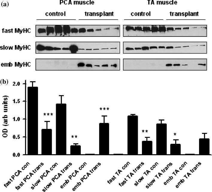
Fig. 6.
Changes in myosin heavy chain protein expression in transplanted muscle. a Western blots of lysates were prepared from the PCA and TA muscles from control animals (con) and transplanted (trans) muscle 7 days following surgery. Samples were electrophoresed and transferred to nitrocellulose membranes as described in “Materials and methods”. Membranes were probed with antibodies directed against fast, slow, or embryonic type MyHC protein. b Graphs show densitometry data as mean ± SEM values (n = 5 animals) *P < 0.05; **P < 0.01; ***P < 0.001 significantly different when transplant muscle is compared with control muscle

