
Figure 4.
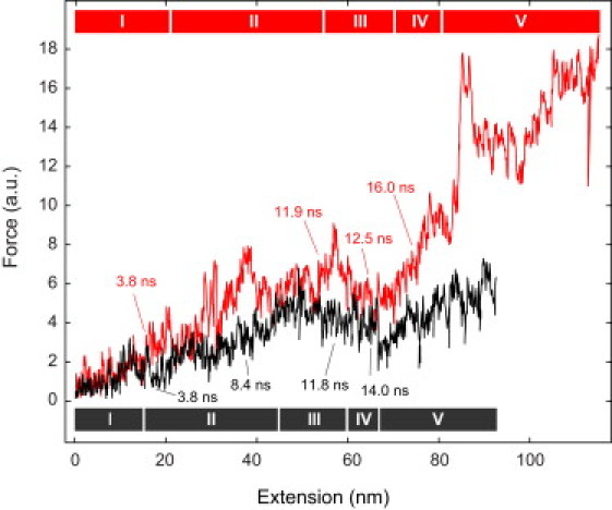
Virtual force spectroscopy of nucleosome unwrapping. From the SMD simulations, force spectroscopy curves were computed. The assignment of the five unwrapping phases and intermediates according to the indicated simulation times corresponds to that given in Fig. 2. The complete nucleosome structure (red line) is shown in comparison to the NUCΔtail structure (black line).