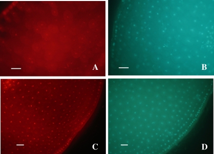
Fig. 6.
Fluorescence micrographs of sunflower seed sections stained with PI (A and C) and DAPI (B and D). Transverse hand-cut sections of an axis corresponding to viable dry seeds (0.04 g H2O g−1 DM) (A and B) and an axis from seeds previously equilibrated at a water content of 0.37 g H2O g−1 DM, and then aged at 35 °C for 7 d (C and D), were stained with PI (5 μg ml−1) and DAPI (300 nM) in PBS medium prior to image capture. Red nuclei correspond to dead cells. Bar=50 μm.

