Fig. 9.
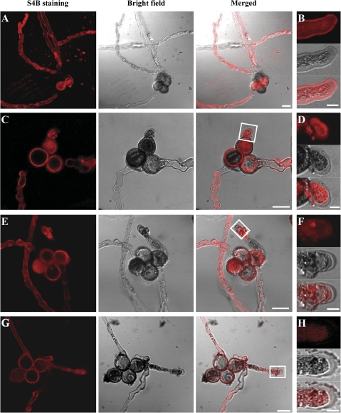
S4B staining of mutant and wild-type pollen tubes. Confocal sections of wild-type qrt1/qrt1 (A, B), csld4-3/+; qrt1/qrt1 (C, D), csld1-1/+; qrt1/qrt1 (E, F), and csld1-1/+; csld4-3/+; qrt1/qrt1 (G, H) pollen tubes stained with S4B. The S4B fluorescence intensity in the cell wall of csld4 (C, D), csld1 (E, F), and csld1 csld4 double mutant (G, H) pollen tubes was much weaker than that of wild-type pollen tubes (A, B). It is noteworthy that the S4B staining is weak at some parts of the pollen tube that are out of the focal planes shown here (especially the pollen tube base near the pollen grain). (B) High magnification image of a wild-type pollen tube showing S4B staining associated with the tube wall and cytoplasm. D, F, and H correspond to the white boxes in C, E, and G, respectively, showing that fluorescence was difficult to detect in the mutant pollen tube wall (five independent observations). Bars: (A, C, E, G) 20 μm; (B, D, F, H) 50 μm.

