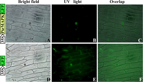
Fig. 4.
Cellular localization of PtrMAPK in onion epidermal cells. PtrMAPK was fused to the N-terminus of green fluorescence protein (PtrMAPK–GFP), which was transformed into onion epidermal cells through Agrobacterium-mediated infection, using GFP as a control. The expression of PtrMAPK–GFP or GFP alone was examined under a universal fluorescence microscope. Bright-field images (A and D), fluorescence images (B and E), and the overlapped images (C and F) of representative cells expressing PtrMAPK–GFP fusion protein (upper panels) or GFP (lower panels) are shown.

