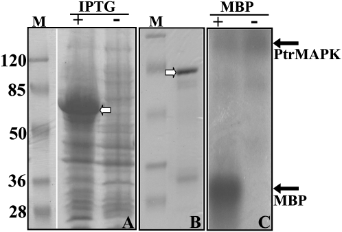
Fig. 5.
Analysis of kinase activity in PtrMAPK. (A) SDS–PAGE separation of protein extracted from E. coli transformed with pGEX-PtrMAPK induced (+) or not (–) with 1 mM IPTG. (B) SDS–PAGE separation of the purified protein derived from the IPTG-induced extract in A. The predicted protein in A and B is shown by an open arrow. M, a molecular weight marker (kDa). (C) Detection of protein phosphorylation. The purified PtrMAPK protein was incubated in a buffer to which MBP for kinase reaction was added (+) or not (–), followed by detection of phosphorylation with SDS–PAGE and autoradiography. The positions of the proteins are shown by the filled arrows.

