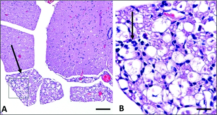
Figure 2.
Histopathology of mice presenting with paresis (mice presenting after 6 mo) (A) Ventral horn, spinal cord. Marked regional axonal degeneration of ventral roots is evident (arrow). Hematoxylin and eosin stain; bar, 50 μm. (B) Ventral root, spinal cord: boxed area from A. Small numbers of mononuclear cells admixed with occasional neutrophils infiltrate degenerate ventral roots (arrow). Hematoxylin and eosin stain; bar, 50 μm.

