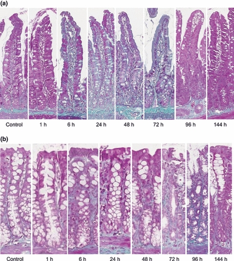
Figure 4.

Masson trichrome staining demonstrating fibrous collagen in the (a) jejunum and (b) colon in a time course model of irinotecan-induced mucositis. Fibrous collagen is stained green on a purple background. Photomicrographs taken at 20× objective.
