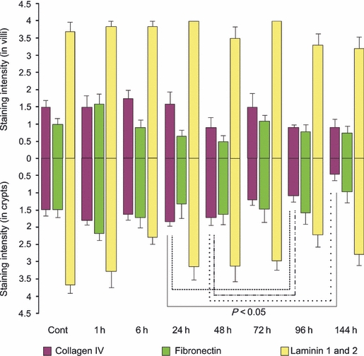
Figure 5.

Extracellular matrix component staining in the jejunum. Top bars illustrate staining in villi while bottom bars illustrate staining in crypts at 1, 6, 24, 48, 72, 96 and 144 h following the administration of 200 mg/kg irinotecan intraperitoneally. All control and experimental tissue were graded by a qualitative scale. The data are means + standard error; significance indicated on the graph.
