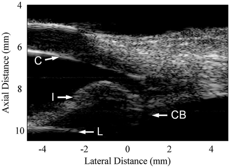
Fig. 11.

Image of an excised bovine eye obtained with the 30-MHz array and custom-designed imaging system. One transmit (6.4 mm) and four receive foci (6.4, 8.75, 9.6, and 11.2 mm) were used to form this image. The cornea (C), iris (I), ciliary body (CB), and the surface of the lens (L) are clearly visible in this image. The displayed dynamic range for this image is 40 dB.
