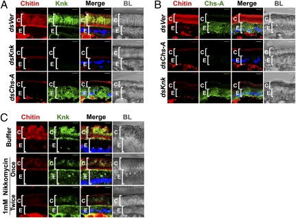
Fig. 3.
TcKnk is mislocalized after disruption of chitin synthesis. (A) Cryosections of T. castaneum pharate adult lateral body walls (20 μm thick) of control TcVer (dsVer), TcKnk (dsKnk), and TcChs-A (dsChs-A) dsRNA-treated insects were immunostained with D. melanogaster Knk antiserum (green). (B) Distribution of TcChs-A was determined by using anti–TcChs-A antibody (green) in various dsRNA-treated insects. Specificities of the D. melanogaster Knk and TcChs-A antibodies as well as the chitin probe were ascertained by using insects subjected to RNAi for these two genes as controls. The punctate distribution of both TcChs-A and TcKnk within cells suggests vesicular localization. (C) Nikkomycin (1 mM, 0.2 μL per insect) was injected either once at the pharate pupal stage or twice (on days 1 and 3 of the pupal stage) before cryosectioning and immunostaining for TcKnk (green) and staining for chitin (red). Chitin, red; proteins, green; DAPI, blue; C, cuticle; E, epithelial cells. (Scale bars: 5 μm.)

