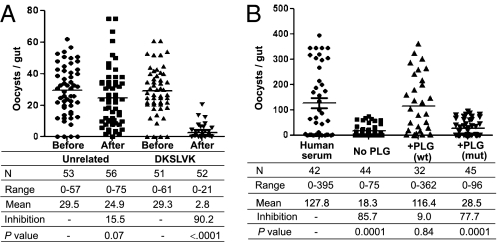
Fig. 5.
Ookinete dependence on its surface enolase and on interaction with mammalian plasminogen for invasion of the mosquito midgut. (A) Inhibition of P. berghei oocyst formation by the DKSLVK peptide. For each experiment, a group of control An. gambiae mosquitoes was fed on a P. berghei-infected mouse (before). The mouse was then injected i.v. with 200 μg of the DKSLVK peptide or of an unrelated control peptide (QPQHFR), and after 10 min, a group of experimental mosquitoes was fed on the same mouse (after). Data for three independent experiments were pooled. N indicates the number of mosquitoes analyzed, range indicates the minimum and maximum number of oocysts per gut, mean indicates the mean number of oocysts per gut, and inhibition indicates the percent decrease of oocyst numbers. The P value was calculated by the Mann–Whitney test. (B) P. falciparum dependence on mammalian plasminogen for oocyst formation. An. gambiae mosquitoes were fed on P. falciparum gametocytes suspended in normal human serum in plasminogen-depleted human serum (No PLG) or in plasminogen-depleted serum with added recombinant WT plasminogen [200 μg/mL; +PLG (wt)] or with added recombinant mutant plasminogen carrying a point mutation in the active site (S741A) [200 μg/mL; +PLG (mut)], as indicated. Data for three independent experiments were pooled.

