
Figure 2.
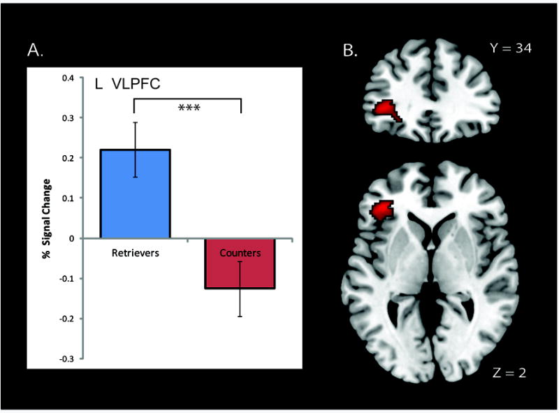
(A) Percentage signal change in the VLPFC across the two groups. (B) Brain areas that showed significant differences in voxel-wise activation levels during the Addition task between Retrievers and Counters. The left ventrolateral prefrontal cortex (VLPFC) showed greater activation in Retrievers compared to Counters. No brain regions showed greater activation in Counters, compared to Retrievers. Three asterisks (***) on graph denote p < .001.