Figure 1.
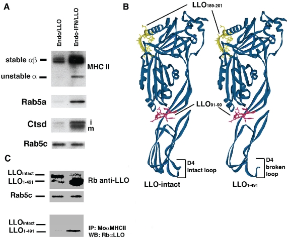
Features of the two LLO structural forms in endosomes. Endosomes were obtained from BM-DM (2 × 10⁸ cells) pre-treated (Endo-IFN/LLO) or not (Endo/LLO) with mIFN-y followed by incubation with 300 µg/ml of LLO. A, endosomal membranes (30 µg) were prepared as described in Materials and Methods and loaded on SDS-PAGE gels, transferred and western-blots developed with rabbit anti-MHC II α chain, rabbit anti-Rab5c, 4F11 monoclonal anti-Rab5a or rabbit anti-Ctsd antibodies. Rab5c is used as control for protein loading. B, 3D model structures represent LLO intact (right structure) and Ctsd-degraded LLO1-491 from (left structure). MHC-I immuno-dominant epitope LLO91-99 is shown in red and MHC-II immuno-dominant epitope LLO189-201 in yellow. C, Upper lane labelled as Rb anti-LLO shows a western-blot of Endo/LLO and Endo-IFN/LLO endosomes that evaluate the levels of LLO intact and Ctsd-degraded LLO1-491 forms in these vesicles. As loading control of these lysates we include Rab5c as in Figure 1A. Lower lanes correspond to immuno-precipates of Endo/LLO and Endo-IFN/LLO endosomes with mouse anti-MHC-II (IAk) (10.3.62 antibody) and western-blot developed with Rb anti-LLO antibody.

