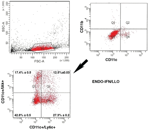
Figure 4.
Analysis of cells recruited to the PEC of animals vaccinated with ENDO-IFN/ LLO vaccine. CBA/J mice were vaccinated i.p with 30 μg of LLO loaded endosomes from IFN-γ activated BM-DM (ENDO-IFN/ LLO) for 7 days (n = 5/vaccination type). Next, mice were inoculated with LM (5 × 10³ bc/mice) for 3 days. PECs were obtained after 2-fold ice-cold washings with Hank's balanced buffer and surface stained with the following FITC or PElabelled antibodies: CD11b (MØ), CD11c (DC) (upper right flow histogram) and CD11c+ cells were gated (Q3 + Q4 regions in plots). Next, PE-CD11c+ cells were analyzed for the percentages of APC-labelled Ly6C or 40F anti-IAk antibodies to distinguish for mature DC (DCm) (CD11c+IAk+) or immature DC (CD11c+Ly6C+) (lower flow histogram). Samples were performed in triplicates and results of the double positive DCm or DCi cells are expressed as the mean ± SD of triplicates (p<0.01).

