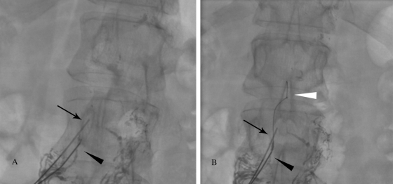
Figure 6.
Fluoroscopic image demonstrating “2-needle” technique. (A) The more superior needle (arrow) compresses the duct, resulting in accumulation of the contrast in the duct below the needle, creating the “target.” The second needle is used to access the duct (black arrowhead). (B) The wire is advanced into the cisterna chyli and thoracic duct (white arrowhead) through the second needle (black arrowhead).

