
Figure 1.
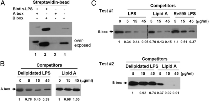
LPS-binding specificity of HMGB1 domains. (A) Biotin-labeled E. coli LPS was incubated with 6× His-tagged HMGB1 A and B box proteins and pull-down assays were performed. The beads were subjected to 12% SDS-PAGE and Western blot analysis was performed using anti-His Ab. (B, C) An aliquot of 5 μg/mL of biotin–LPS was incubated with 5 μg/mL of His-tagged A box or B box HMGB1 protein that had been preincubated with various amounts of E. coli delipidated LPS, S. minnesota lipid A, S. minnesota Re595 LPS, or WT S. minnesota LPS as inhibitors. Biotin–LPS was precipitated and analyzed using Western blotting with an anti-His Ab. (C) The line indicates the cutline of the same blot membrane. Data shown are representative of two independent experiments.