
Figure 2.
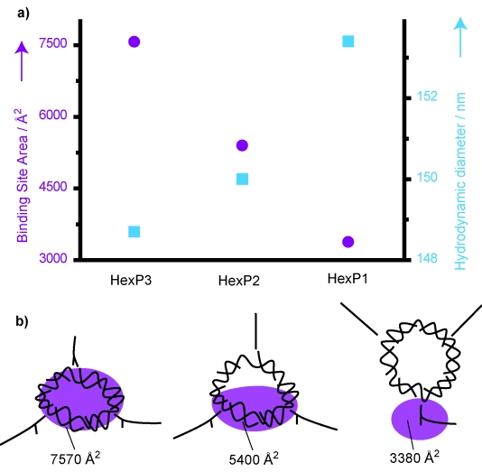
a) The binding site area of the hexagonal-like 2D DNA (purple circles) and the diameter of the liposome–DNA construct (blue squares) are shown as a function of the number of anchoring points. The diameter of liposomes in the absence of DNA was 130 nm. b) An interpretation of the data is shown: whereas HexP3 is lying flat on the surface, HexP1 is more perpendicular to the surface, and HexP2 is somewhere in between.