
Fig. 5.
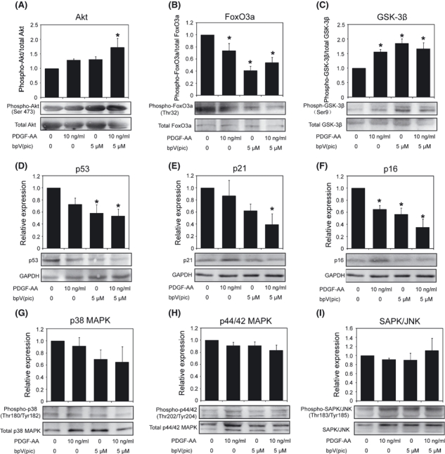
Western blot assay of relevant signal molecules in human foreskin-derived skin-derived precursors (hSKPs) cultured for 12 days in different conditions. (A–C) Phosphorylation of Akt, FoxO3, and GSK-3β in hSKPs cultured without (control) or with PDGF-AA, bpV(pic), and their combination. (D–F) Relative expression of p53, p21, and p16 in hSKPs cultured without (control) or with PDGF-AA, bpV(pic), and their combination. (G–I) Phosphorylation of p38 MAPK, p44/42 MAPK, and SAPK/JNK in hSKPs cultured without (control) or with PDGF-AA, bpV(pic), and their combination. Histograms show statistical results of gray-scale analysis of three independent cultures and representative immunoblots are attached below. Asterisk means P < 0.05 when compared with control.