Figure 4.
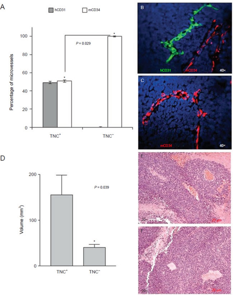
Orthotopic NB tumors formed by TNC+, but not TNC− cells, from the HTLA-230 cell line contain endothelial microvessels lined by tumor-derived endothelial cells. TNC+ cells were isolated from HTLA-230 NB cells by magnetic cell sorting using anti-TNC mAb. TNC-enriched and TNC-depleted cells were injected orthotopically in nude mice. (A) Human (grey columns) and murine (white columns) microvessels in orthotopic tumors formed by TNC+ and TNC− cells in immunodeficient mice. Columns represent mean percent values from 12 different tumors; bars represent SD (P = 0.029; Mann-Whitney test). (B) Double immunofluorescence staining of ortothopic tumors formed by TNC+ cells with hCD31 (green) and mCD34 (red) mAbs. (C) Double immunofluorescence staining of ortothopic tumors formed by TNC− cells with hCD31 (green) and mCD34 (red) mAbs. Nuclei are stained with DAPI (blue). (B, C) Original magnification 40×. (D) Quantification of the volume of tumors formed by TNC+ and TNC− cells demonstrated that TNC+ cells formed significantly larger tumors than TNC− cells (*P = 0.039). (E) Hematoxylin/eosin staining of orthotopic tumor formed by TNC+ cells. (F) Hematoxylin/eosin staining of orthotopic tumor formed by TNC− cells. (E, F) Original magnification 20×.

