Figure 5.
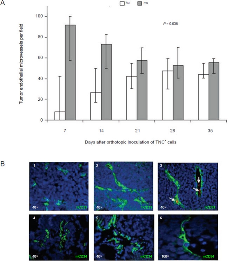
Kinetics of human and mouse endothelial microvessel formation in orthotopic tumors formed by TNC+ cells in immunodeficient mice. (A) Kinetics of hCD31+ (white columns) and mCD34+ (grey columns) tumor endothelial microvessels per field (expressed as median percentages from 20 different tumors) from 7 to 35 days after orthotopic inoculation of TNC+ cells isolated from HTLA-230 cell line. (B) (1) On day 7, CD31+ human endothelial cells appear as small clusters, as assessed by immunofluorescence. (2-3) From day 14 human endothelial microvessels display mature morphology. Erythrocytes are visible in the lumen of most human microvessels (arrows). (4) On day 7, CD34+ mouse endothelial cells lined nascent endothelial microvessels. (5-6) From day 14 mouse endothelial microvessels display mature morphology. Nuclei are stained with DAPI (blue). Original magnification 100× in B6; 40× in all the other panels.

