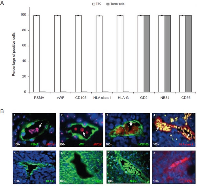
Figure 6.
Immunophenotypic characterization of human endothelial microvessels in orthotopic tumors formed by TNC+ NB cells in immunodeficient mice. (A) Immunofluorescence analyses show the expression of PSMA, von Willebrand factor (vWF), CD105, HLA-class I, HLA-G, GD2, NB84 and CD56 in TEC and in tumor cells. Columns represent mean values obtained from the count of 20 fields/slide in 10 different orthotopic NB tumors formed by TNC+ cells; bars represent SD. (B) (1-2) Immunophenotype of human endothelial microvessels as assessed by immunofluorescence combined with MYCN FISH. (1) Tissue sections were stained with anti-PSMA (green), (2) anti-vWF (green), (3) anti-hCD105 (green), (4) anti-HLA-class I, (5) anti-HLAG, (6) anti-GD2, (7) anti-NB84, and (8) anti-CD56. Nuclei are stained with DAPI (blue). Original magnification 40× in B5 and 100× in other images.

