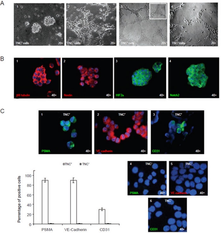
Figure 7.
Plasticity of TNC+ cells isolated from the HTLA-230 cell line. (A) (1) TNC+ cells enriched from the HTLA-230 NB cell line and cultured in serum-free medium supplemented with bFGF and EGF form neurospheres in vitro. (2) TNC− cells grow as an adherent layer in serum-free medium supplemented with bFGF and EGF as parental cell line. (3) Representative micrograph of the complete network of tubes formed by TNC+ cells (1-3 × 10⁴ cells/well) plated on growth factor-reduced Matrigel after stimulation with VEGF (5-15 ng/ml) and bFGF (20-50 ng/ml) after 6 h. Tubes of the TNC+ cells persisted for up to 5 days. The inset shows an enlargement of a detailed network of tubes. (4) Representative micrograph of the weak network of tubes formed by TNC− cells (1-3 × 10⁴ cells/well) plated on Matrigel. Structures of TNC− cells rapidly dissolved in a few hours. (B) (1) Neurospheres express βIII tubulin (red), (2) nestin (red), (3) HIF-2α (green), (4) Notch 2 (green), as assessed by immunofluorescence with specific mAbs. (C) (1) TNC+ cells cultured in differentiating medium supplemented with 2% FCS and VEGF for 10-15 days acquire expression of the endothelial-specific markers PSMA, (2) VE-Cadherin, and (3) CD31. (4-6) TNC− cells did not express the markers. (4), (5) and (6) show negative staining for PSMA, VE-Cadherin and CD31 of TNC− cells cultured in endothelial differentiation medium. Nuclei are stained with DAPI (blue). Original magnification 40×. Lower left panel, PSMA and VE-Cadherin were upregulated in more than 90% of the differentiating TNC+ cells, while CD31 was detected on approximately 30% of them. Columns (TNC+ white columns and TNC− grey columns) represent mean values from five replicate experiments; bars represent SD.

