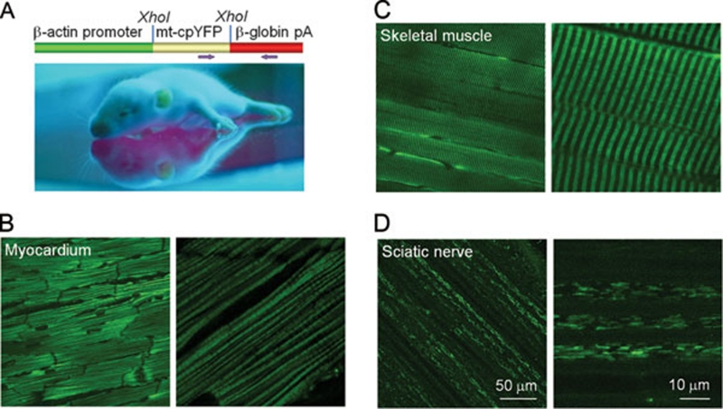
Figure 1.
Pan-tissue expression of mt-cpYFP in transgenic mice. (A) A neonatal mt-cpYFP transgenic mouse under UV illumination. Top: plasmid construct for generating the transgenic mouse. The full-length mt-cpYFP DNA was cloned into pUC-CAGGS vector downstream of the chicken β-actin promoter. Arrows indicate the positions of forward and backward primers for genotyping. (B-D) Confocal images of mt-cpYFP fluorescence in myocardium (B), gastrocnemius muscle (C) and sciatic nerve trunk (D) at low and high magnifications. Note the cell type-specific pattern of intracellular mitochondrial distribution. (B-D) use the same scale bar in D.

