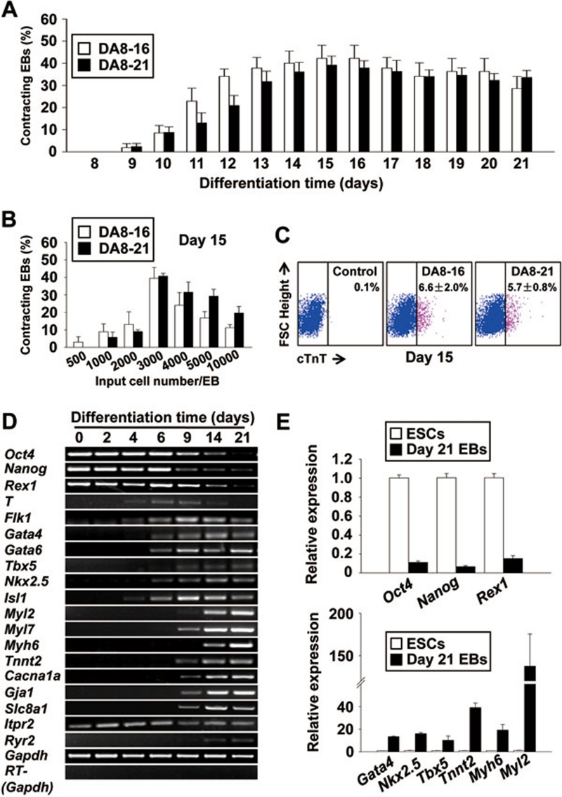
Figure 3.
In vitro differentiation of the rESCs induces expression of mesoderm, cardiac progenitor markers, cardiomyocyte markers, and development of contracting EBs over time. (A) The percentage of EBs with contracting areas during differentiation. A total of 222 EBs from clone DA8-16 and 292 EBs from clone DA8-21 were counted from three independent experiments. (B) Identification of the optimal input cell number in each EB for cardiac differentiation of rESCs. Data were analyzed from three independent experiments. (C) Flow cytometry analysis showing the percentage of cardiomyocytes in the total population derived from both rESC lines. Results were quantified at differentiation day 15 from three independent assays. (D) RT-PCR analysis showing the downregulation of pluripotency marker expressions and the upregulation of mesoderm, cardiac progenitor, and cardiac marker expressions. (E) Q-PCR analysis of gene expression levels for pluripotency (upper panel), cardiac progenitor and cardiomyocyte markers (lower panel) in undifferentiated rESC and in day-21 EBs. Data were collected from three independent experiments.

