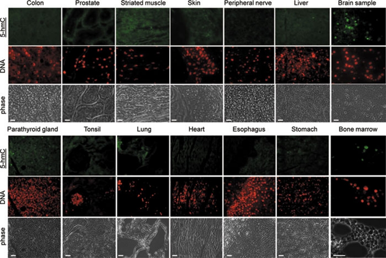
Figure 5.
The presence of immunochemically detectable 5-hydroxymethylcytosine (5-hmC) levels is restricted to bone marrow and neuronal tissues in adult mammals. A range of indicated normal adult human tissues from a Folio histological tissue array were immunostained with anti-5-hmC antibody. Phase views are shown. An identical maximal exposure of the images made using the 488 nm (5-hmC, tyramide) filter is shown for all the samples, except brain and bone marrow, to illustrate the pattern of background staining, which does not correspond to the DNA signal (indicated) in the majority of tissues. All the triplicate tissue samples on the array exhibited identical staining patterns. Six different bone marrow specimens were tested and gave essentially the same results. Representative views are shown. Scale bars are 20 μm, except 30 μm in bone marrow sample. Essentially the same results were obtained with samples of ovary, testis, kidney, small intestine, salivary gland, uterus, uterine cervix, mesothelium, eye muscle, adrenal gland, pancreas and breast tissue (see Supplementary information, Figure S9). The experiments were performed using the Diagenode antibody.

