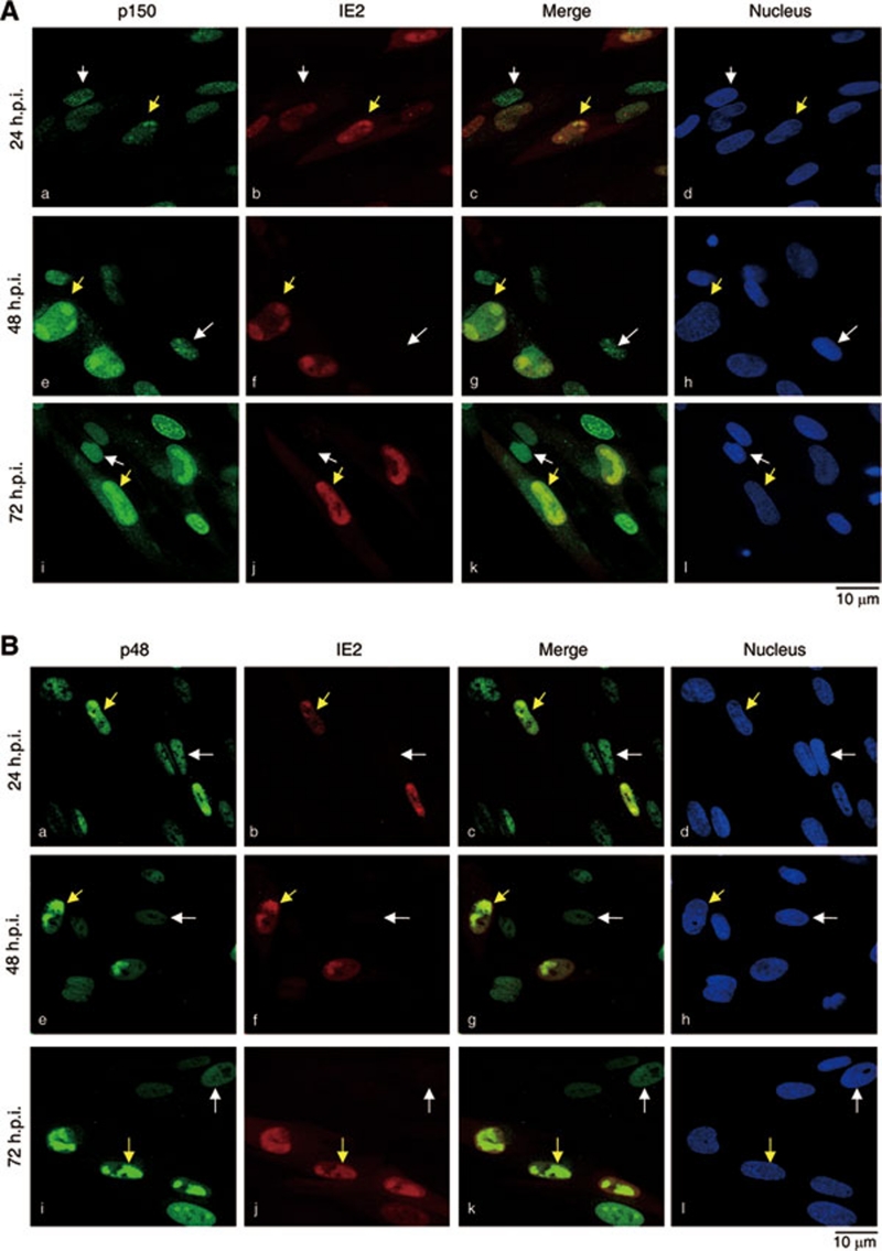
Figure 3.
CAF1 colocalizes with IE2 in specific nuclear compartments in HCMV-infected cells. MRC-5 cells were serum starved for 72 h and infected with HCMV at an MOI of 0.5, followed by incubation with serum-containing medium. At the indicated h.p.i., the localization of p150 and IE2 (A), and p48 and IE2 (B) was examined by double staining with corresponding antibodies. Images were obtained from Leica TCS NT confocal microscopy. White and yellow arrowheads indicate the uninfected and infected cells, respectively, based on the expression of HCMV IE2. The 4′, 6-diamidino-2-phenylindole dihydrochloride was used to stain the nucleus. All shown images represent the phenomenon occurring in more than 90% of the population. The exception has been indicated in main text.

