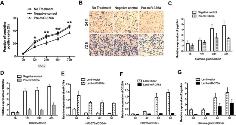
Figure 2.
Over-expression of miR-376a inhibits the erythroid differentiation of K562 cells and CD34+ HPCs. (A) Benzidine staining of K562 cells that were untransfected, or transfected with a negative control precursor (negative control) or a miR-376a precursor (Pre-miR-376a) were induced with hemin for 12, 24, 48, and 72 h. Pre-miR-376a reduces the percentage of benzidine-positive cells at different time points. The fraction of benzidine-positive cells was determined by a benzidine cytochemical test. Error bars represent the standard deviation and were obtained from three independent experiments. * indicates P < 0.05, ** indicates P < 0.01. (B) Representative benzidine staining of un-transfected and transfected K562 cells following 24 and 72 h of hemin induction. (C-D) Quantitative real-time PCR analysis of γ-globin and CD235A in the transfected K562 cells after hemin induction for 12, 24, and 48 h. Real-time PCR analysis was performed in triplicate and the expression levels of γ-globin and CD235A mRNAs were normalized to GAPDH mRNA. Error bars represent standard deviation. (E) Quantitative real-time PCR analysis of miR-376a in the CD34+ HPCs that were infected with a lenti-virus expressing mature miR-376a (lenti-miR-376a) or a control virus (lenti-vector) and cultured in erythroid induction medium for the indicated time. (F-G) Quantitative real-time PCR analysis of γ-globin and CD235A in the CD34+ HPCs infected with the recombinant viruses and cultured in erythroid induction medium for the indicated time.

