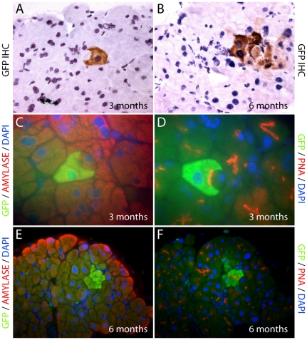
Figure 2. Normal pancreas in bone marrow transplant recipients.
GFP immunohistochemistry of pancreata identified individual GFP+ve donor-derived cells with acinar cell morphology from 3 months post transplantation (A), and occasional entire acinar units (B), were observed from 6 months post transplantation. Immunofluorescence images of donor derived GFP+ve acinar cells co-staining positive for both GFP and amylase (C, E); and GFP and PNA (D, F).

