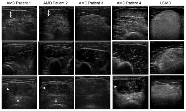
Figure 2. Patterns of Muscle Pathology on Ultrasound in Acid Maltase Disease.
Ultrasound of the biceps brachii and brachialis (Row 1), triceps brachii (Row 2) and rectus femoris and vastus intermedius (Row 3) is shown for four patients with acid-maltase disease (AMD) aged 45–63 years and a 49 year-old woman with limb-girdle muscular dystrophy (LGMD). In AMD patients, there is sparing of the triceps brachii compared to the biceps brachii (patients 1–4), sparing of the superficial portion of the biceps brachii (arrows, patients 1 and 2), and sparing of the rectus femoris (arrowhead) compared to the vastus intermedius (*, patients 1, 2, and 4). In contrast, in LGMD there is more diffuse, homogenous involvement of the proximal arm and leg muscles.

