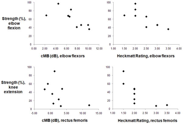
Figure 3. Ultrasound in adults with AMD: Relations to Strength.
Elbow flexion strength (top, expressed as a percent of normal) decreased with both higher quantitative (cMB, rs=−0.8, p=0.008) and qualitative (Heckmatt rating, rs=−0.8, p=0.004) ultrasound measures of muscle pathology in the elbow flexors. Knee extension strength decreased with higher Heckmatt ratings (rs=−0.9, p=0.001) but not cMB levels (rs =−0.4, p=0.2) of the rectus femoris. Compared to the elbow flexors, ultrasound measurements of the rectus femoris were relatively insensitive to changes in strength. For example, five subjects had a Heckmatt rating of two despite differences in knee extension strength from 48 to 13% of normal.

