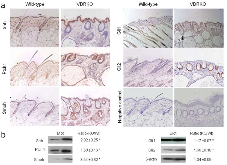
Figure 2. Increased protein levels of Hh signaling pathway members in VDRKO mice.

A. Shh, Ptch1, Smoh, Gli1 and Gli2 proteins as shown by the brown signal were detected in the epidermis and hair follicles from VDRKO mice and their wild-type littermates at 11 weeks after birth by immunocytochemistry. Slides were counterstained with hematoxylin (blue stain). The bar denotes 50 μm. B. Shh, Ptch1, Smoh, Gli1 and Gli2 protein levels were quantified by western blot in skin from VDRKO mice and their wild-type littermates and compared to beta-actin as a control. The numerical value represents the average ratio of VDRKO band intensity versus wild-type band intensity after subtraction of background level from three independent experiments. * p<0.05.
